Tel/Whatsapp: +86 19352847010
Email: [email protected]

- Product
Ultrasound System
Life Information Monitoring System
Infusion & Syringe System
- About Us
- Media & Events
- Service
- Contact Us
Tel/Whatsapp: +86 19352847010
Email: [email protected]
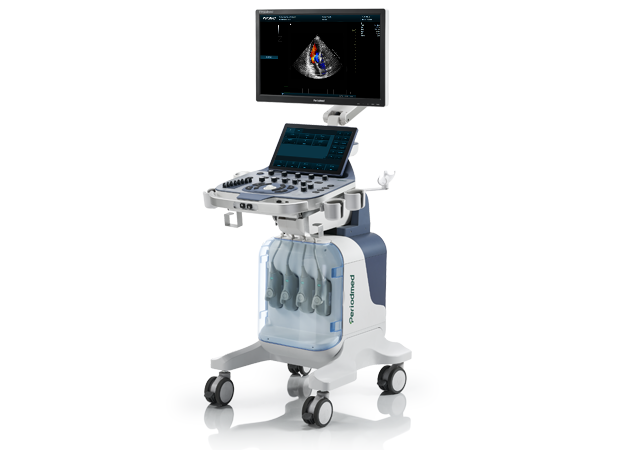
High-End MSKUS Rheumatology|Revo T2

As a professional advanced MSK ultrasound rheumatology system, Revo T2 adopts multi-beam synthesis and high-precision beamforming technology. It delivers ultra-fine musculoskeletal tissue imaging tailored for rheumatic disease ultrasound monitoring. Optimized for complex joint structures, it supports full quantitative evaluation of rheumatic lesions, laying a reliable imaging foundation for long-term rheumatology clinical follow-up and condition grading.
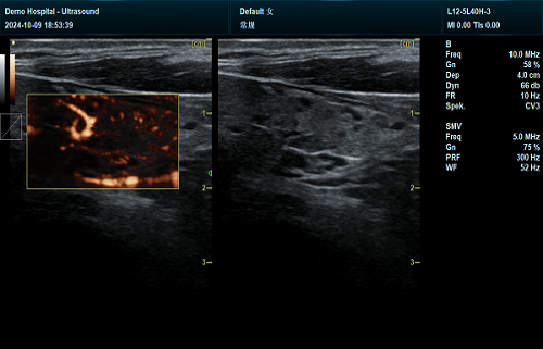
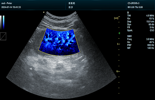
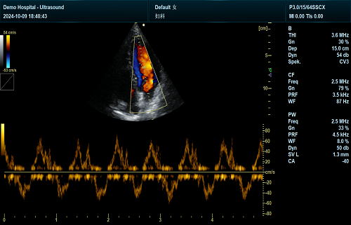
Revo T2 features superb microvascular imaging rheumatoid arthritis activity detection, realizing sensitive synovitis microvascular imaging invisible on conventional Doppler. It captures subtle microvascular hyperplasia in inflamed synovium, accurately reflecting early inflammatory activity. This advanced modality solves the limitations of traditional imaging, enabling early identification of subclinical inflammation for timely clinical intervention.
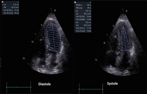
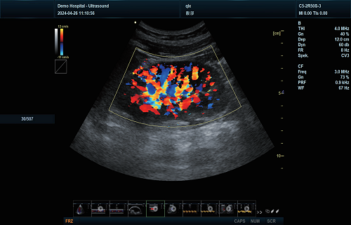
Supporting rheumatoid arthritis Doppler quantification and ultrasound synovitis erosion quantification, the system provides objective numerical data for joint inflammation assessment. It precisely identifies synovial hypertrophy, bone erosion and exudation, achieving accurate ultrasound joint damage progression rheumatology tracking. Clinicians rely on standardized quantitative indicators to judge disease activity and adjust treatment regimens scientifically.
Equipped with tissue harmonic imaging, spatial compound imaging and intelligent speckle reduction, Revo T2 optimizes joint and soft tissue image clarity. Its one-click intelligent image calibration and multi-angle echo fusion eliminate imaging artifacts. Rich professional measurement packages support full-cycle quantitative monitoring of rheumatic joints, ensuring stable and consistent diagnostic results for repeated follow-up scans.
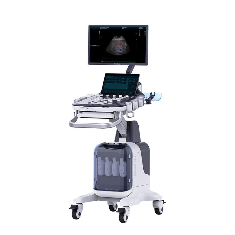
Configured with a 23.8-inch HD monitor and 13.3-inch adjustable touch screen, Revo T2 presents intuitive high-resolution joint imaging. Four full-active probe channels adapt to multi-site joint scanning. Supporting DICOM system linkage and multi-language operation, it standardizes rheumatic disease data archiving, perfectly matching long-term quantitative monitoring needs of professional rheumatology clinics.
| Application | |||
| Generic | Obstetrics | Gynecology | Abdomen |
| Vascular | Small Parts | Urology | Thyroid |
| Cardiology | MSK | ||
| Image Formats | |||
| AVI | BMP | JPG | PNG |
| DICOM | |||
| Transducer Types | |||
| Convex probe | Linear probe | Phased array | |
| Volume array | Intracavity probe | Micro convex array | |
| Operating Mode | |||
| B | 2B | 4B | CW |
| M | CF | B/M | PW |
| PDI | PIP | Trapezoid | Panorama |
| 3D/4D(Optional) | Wide view (Optional) | Picture in Picture | Color M |
| Anatomy M | Directional Power Doppler Imaging | TDI(Optional) | Needle Enhance (Optional) |
Contrast Enhance(Optional) | Elastography (Optional) | SMV(Optional) | |
| System Architecture | |||
| Elements: 64/128/160/192 | Frequency Range: 2.0-22.0 MHz | Memory:8G | Hard Drive:256G |
| Dynamic Range: ≥120db | 13.3 Inches touch screen | Resolution:1920*1080 | Beam forming 16 |
| Frequency Range: 2.0-22.0 MHz | 23.8 Inches High Resolution LED Monitor | ||
| Physical Specs | |||
| 23.8 Inches High Resolution LED Monitor | Magnetic latch closure | Built-in speaker | Brightness and contrast adjustable |
| Languages | |||
| English | Arabic | French | Indonesian |
| Italian | Portuguese | Spanish | Chinese |
| German | Russian | ||


 EN
EN CN
CN