Tel/Whatsapp: +86 19352847010
Email: [email protected]

- Product
Ultrasound System
Life Information Monitoring System
Infusion & Syringe System
- About Us
- Media & Events
- Service
- Contact Us
Tel/Whatsapp: +86 19352847010
Email: [email protected]
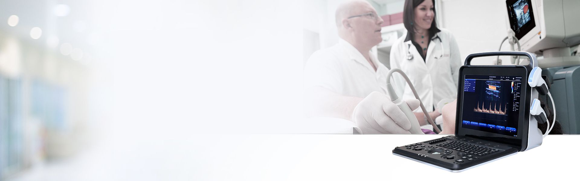
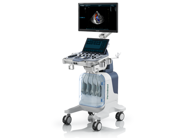
Advanced Elastography Sports Medicine | Revo T2

Revo T2 integrates professional elastography sports medicine technology tailored for elite athlete musculoskeletal evaluation. Equipped with multi-beam synthesis and high-precision beamforming algorithms, it delivers stable and detailed basketball tendon ultrasound imaging. It supports shear wave elastography basketball analysis, enabling quantitative tendon stiffness detection for systematic sports tendon health screening in basketball training and medical management.
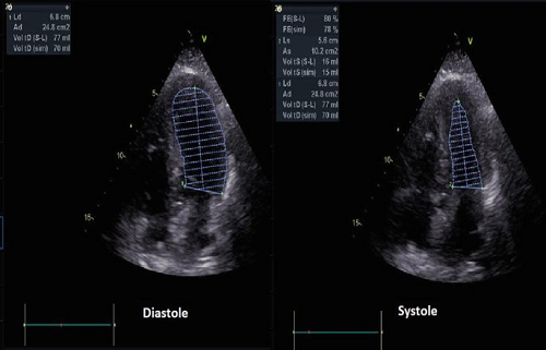
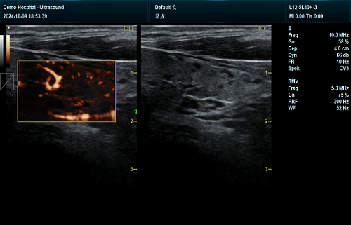
Specialized for SWE Achilles tendon basketball players assessment, Revo T2 accurately measures Achilles tendon stiffness athletes data. Its intelligent sound speed correction and multi-angle fusion imaging capture subtle tendon structural changes caused by repeated jumping and sprinting. Clinicians conduct precise injury risk screening, identifying early degenerative lesions that conventional B-mode ultrasound cannot detect for proactive athlete protection.
Revo T2 provides professional patellar tendon ultrasound elite scanning for basketball athletes’ knee tendon evaluation. It enables real-time ultrasound tendon stiffness monitoring training load, tracking tendon fatigue and cumulative strain during intensive training. This quantitative assessment effectively assists patellar tendinopathy ultrasound return to sport judgment, offering objective data for scientific training adjustment and injury prevention.
Built with dedicated MSK examination mode and full intelligent puncture enhancement, Revo T2 optimizes imaging for superficial tendon tissues. Intelligent speckle reduction and one-click image optimization ensure clear, consistent tendon images during repeated athlete screenings. Multiple advanced imaging modes support comprehensive soft tissue evaluation, improving accuracy for sports tendon lesion diagnosis and rehabilitation monitoring.
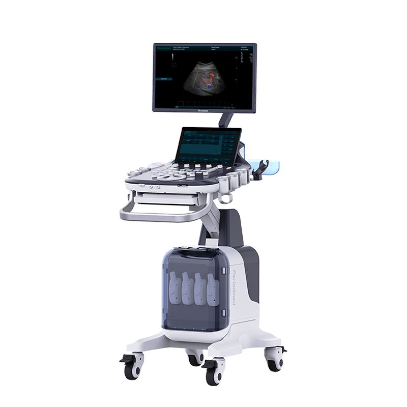
Featuring a 23.8-inch HD monitor and 13.3-inch adjustable touch screen, Revo T2 delivers ultra-clear dynamic tendon imaging. Four fully activated probe channels support fast switching for Achilles and patellar tendon scans. With complete sports measurement packages and DICOM compatibility, it standardizes tendon data recording, perfectly serving elite team sports medicine and professional athlete return-to-sport assessment.
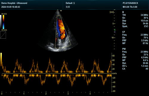
| Application | |||
| Generic | Obstetrics | Gynecology | Abdomen |
| Vascular | Small Parts | Urology | Thyroid |
| Cardiology | MSK | ||
| Image Formats | |||
| AVI | BMP | JPG | PNG |
| DICOM | |||
| Transducer Types | |||
| Convex probe | Linear probe | Phased array | |
| Volume array | Intracavity probe | Micro convex array | |
| Operating Mode | |||
| B | 2B | 4B | CW |
| M | CF | B/M | PW |
| PDI | PIP | Trapezoid | Panorama |
| 3D/4D(Optional) | Wide view (Optional) | Picture in Picture | Color M |
| Anatomy M | Directional Power Doppler Imaging | TDI(Optional) | Needle Enhance (Optional) |
Contrast Enhance(Optional) | Elastography (Optional) | SMV(Optional) | |
| System Architecture | |||
| Elements: 64/128/160/192 | Frequency Range: 2.0-22.0 MHz | Memory:8G | Hard Drive:256G |
| Dynamic Range: ≥120db | 13.3 Inches touch screen | Resolution:1920*1080 | Beam forming 16 |
| Frequency Range: 2.0-22.0 MHz | 23.8 Inches High Resolution LED Monitor | ||
| Physical Specs | |||
| 23.8 Inches High Resolution LED Monitor | Magnetic latch closure | Built-in speaker | Brightness and contrast adjustable |
| Languages | |||
| English | Arabic | French | Indonesian |
| Italian | Portuguese | Spanish | Chinese |
| German | Russian | ||


 EN
EN CN
CN