EN
EN CN
CN
Linear Ultrasound Probes vs. Convex Ultrasound Probes
For global healthcare distributors, understanding the nuances of ultrasound probes is critical to meeting clinical needs across specialties. Ultrasound probes— the "eyes" of diagnostic imaging—determine the accuracy, depth, and scope of scans, directly impacting patient care. This guide breaks down the core differences between linear and convex probes, their specialized applications, and how Periodmed’s wireless handheld ultrasound devices leverage these probe technologies to deliver unmatched value to clinics, hospitals, and mobile care settings worldwide.
What Is an Ultrasound Probe?
An ultrasound probe (transducer) is a core component of diagnostic ultrasound systems, responsible for generating and receiving high-frequency sound waves (ultrasound) that interact with bodily tissues. When sound waves bounce back (echoes), the probe converts these signals into digital images. Beyond imaging, probes dictate:
● Examined body position: Probe design (e.g., shape, frequency) determines which tissues (superficial vs. deep) can be scanned.
● Imaging procedure scope: Specialized probes enable targeted exams (e.g., vascular access, fetal monitoring) by optimizing wave penetration and resolution.
Defining Linear vs. Convex Ultrasound Probes
While both probes serve diagnostic purposes, their design and technology are engineered for distinct clinical needs.
● Linear Ultrasound Probe
A linear probe features a straight, rectangular transducer array that emits sound waves in parallel. It is designed to image superficial, linear structures (e.g., blood vessels, nerves, soft tissues) where high detail is critical.
● Convex Ultrasound Probe
A convex probe (also called a curved array probe) has a curved transducer array that emits sound waves in a fan-like pattern. It is optimized for deep-tissue imaging, as the curved design expands the field of view (FOV) at greater depths, making it ideal for organs and large anatomical regions.
Key Differences of Linear and Convex Probes
The choice between linear and convex probes hinges on clinical goals—resolution vs. depth, superficial vs. deep tissues. Below is a detailed comparison:

Core Applications
Linear
● Vascular access (central line placement, IV insertion), Thyroid/soft tissue imaging, Musculoskeletal (MSK) exams (e.g., sports injuries, joint evaluations), Nerve blocks/regional anesthesia
vs Convex
● Abdominal FAST (Focused Assessment with Sonography for Trauma) exams, OB/GYN assessments (fetal monitoring, pelvic scans), Trauma scans (deep organ evaluation), Renal (kidney) and aortic evaluations
Key Characteristics
Linear
● High frequency (5.0–12.0 MHz): Delivers exceptional resolution for fine structures (e.g., nerve bundles, thyroid nodules).
● Rectangular FOV: Produces flat, clear images of superficial anatomy (e.g., blood vessel walls).
● Limited depth: Typically effective up to 4–5 cm; not suitable for deep organs (e.g., liver, uterus).
Movement sensitivity: Requires steady handling to avoid image artifacts.
Convex
● Mid-to-low frequency (2.5–6.0 MHz): Prioritizes depth over ultra-high resolution, penetrating 10–20 cm to reach deep organs.
● Fan-shaped FOV: Expands at depth, covering large areas (e.g., the entire abdomen) in fewer scans.
● Depth versatility: Ideal for abdominal, pelvic, and fetal imaging.
● Robust to movement: More forgiving in dynamic settings (e.g., trauma resuscitation).
Beyond Linear & Convex: 5 Other Commonly Used Probes
While linear and convex probes cover most primary needs, specialized scenarios require additional probe types:
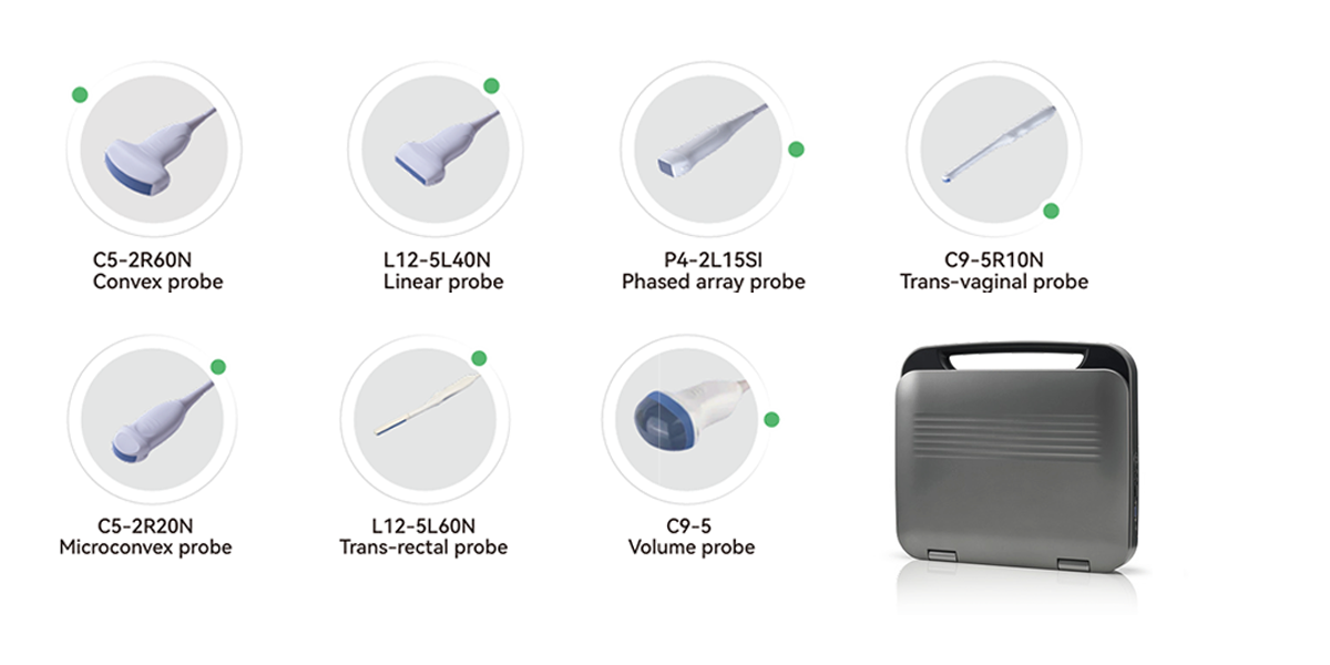
● Phased Array Probes: Small, compact design with a narrow FOV; used for cardiac imaging (e.g., echocardiograms) in tight spaces.
● Transvaginal Probes: High-frequency, endocavitary probes for detailed OB/GYN imaging (e.g., early pregnancy scans).
● 3D/4D Volume Probes: Capture volumetric data for dynamic imaging (e.g., fetal movement in 4D).
● Trans-Rectal Probes: Used for prostate exams and male pelvic imaging.
● Micro-Convex Probes: Miniaturized convex probes for pediatric or small-animal imaging, balancing depth and FOV. Learn more about Periodmed ultrasound probes.
Wireless Handheld Ultrasound Solutions with Various Probes
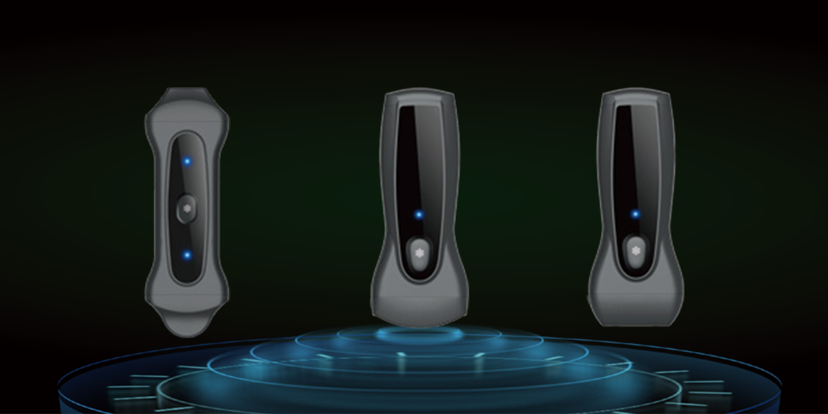
Periodmed’s Aura series is engineered to align with the unique strengths of linear, convex, and dual-probe technologies, delivering portability, durability, and clinical precision—ideal for global distribution to diverse healthcare settings (clinics, rural hospitals, mobile care).
1. Dual Probe (Linear + Convex): Aura PIU2B
Designed for pediatric care (and beyond), the PIU2B integrates both linear and convex probes to cover superficial and deep-tissue needs:
● Linear probe (5.0–12.0 MHz): Excels in MSK exams (joint evaluations) and soft tissue imaging (e.g., pediatric thyroid).
● Convex probe (2.5–6.0 MHz): Ideal for abdominal scans and fetal monitoring in neonates.
● Key Advantages: 212g ultra-lightweight design (pocket-sized), IPX7 waterproofing (easy disinfection, reduced cross-infection risk), speckle reduction + harmonic imaging (clearer anatomical details), and 3200mAh battery (60-minute full charge). Supports 10+ languages (English, Chinese, Spanish, etc.) for global use.
2. Linear Probe: Aura PAU1A
The PAU1A is a dedicated linear probe device optimized for MSK, vascular, and soft tissue imaging—perfect for orthopedics, pain management, and emergency care:
● Linear probe specs: 5.0–12.0 MHz frequency (high resolution), 20–150 mm scanning depth, 128-element channel (consistent imaging).
● Key Advantages: Wireless connectivity (Android/iOS/Windows), protective case with built-in wireless charger, and 212g weight (reduces clinician fatigue during prolonged use). Ideal for sports injury clinics, mobile vascular services, and anesthesia departments.
3. Convex Probe: Aura PAU1B
The PAU1B is a convex probe specialist for abdominal, OB/GYN, and deep-organ imaging—critical for primary care, rural hospitals, and prenatal clinics:
● Convex probe specs: 2.5–6.0 MHz frequency (deep penetration), 40–300 mm scanning depth, 128-element channel.
● Key Advantages: Harmonic Imaging Technology (THI) (boosts contrast for subtle abnormalities), IPX7 waterproofing, and ergonomic design (enhances patient comfort during pelvic/abdominal scans). Supports DICOM storage for seamless integration with hospital systems.
Partner With us Now
As a global distributor, you need products that adapt to diverse clinical needs—from pediatric care to MSK clinics, from rural OB/GYN services to emergency trauma care. Contact us today to discuss partnership opportunities. Together, we’ll bring cutting-edge ultrasound technology to clinics and patients worldwide.