Tel/Whatsapp: +86 19352847010
Email: [email protected]

- Product
Ultrasound System
Life Information Monitoring System
Infusion & Syringe System
- About Us
- Media & Events
- Service
- Contact Us
Tel/Whatsapp: +86 19352847010
Email: [email protected]
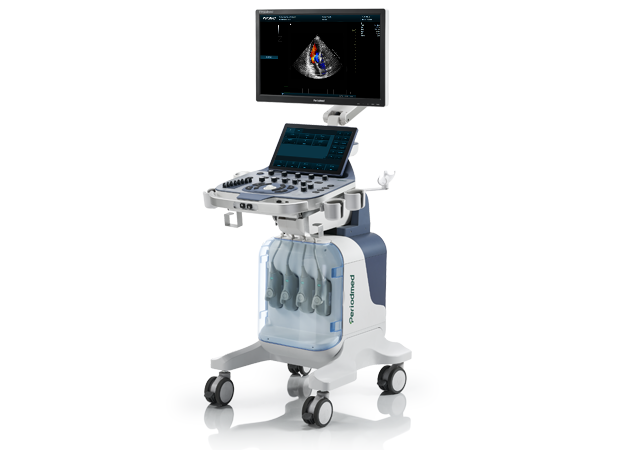
Advanced Structural Heart Ultrasound |Revo T2

Revo T2 trolley ultrasound system delivers premium structural heart ultrasound performance tailored for complex interventional cardiology procedures. Powered by next-generation beam compounding and sound velocity calibration technology, it eliminates imaging artifacts and boosts focusing accuracy. It supports reliable intraprocedural cardiac ultrasound visualization, laying a solid foundation for precise structural heart intervention and intraoperative monitoring.
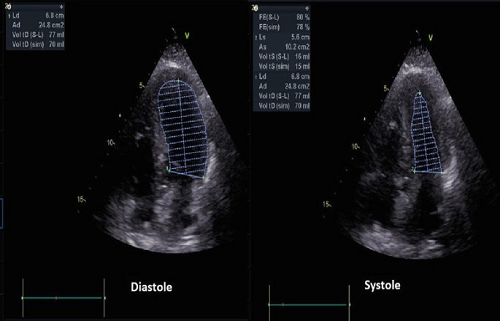
Equipped with professional 4D echocardiography intervention capabilities, Revo T2 provides immersive spatial cardiac anatomy visualization. Its high-sensitivity transducer and GPU-accelerated algorithm capture dynamic 3D/4D cardiac motion in real time. As core 4D cardiac imaging interventional cardiology equipment, it solves the limitations of traditional 2D imaging for complex structural heart lesion assessment.
Revo T2 excels in real-time 3D ultrasound for LAA closure, offering intuitive panoramic views of the left atrial appendage morphology and spatial structure. Dynamic multi-angle imaging assists clinicians in accurate sizing, device positioning and post-closure effect verification. It effectively reduces operational deviations, ensuring safer and more standardized ultrasound guided cardiac procedure for LAA occlusion interventions.
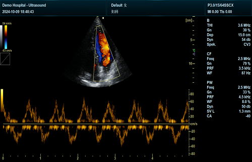
Optimized for handheld ultrasound TAVR guidance, the system supports full-process monitoring for transcatheter aortic valve replacement surgeries. High-definition color Doppler and tissue imaging clearly display valve movement and blood flow changes. It provides real-time intraoperative feedback for prosthesis alignment and fitting evaluation, improving accuracy and safety for all valvular interventional operations.
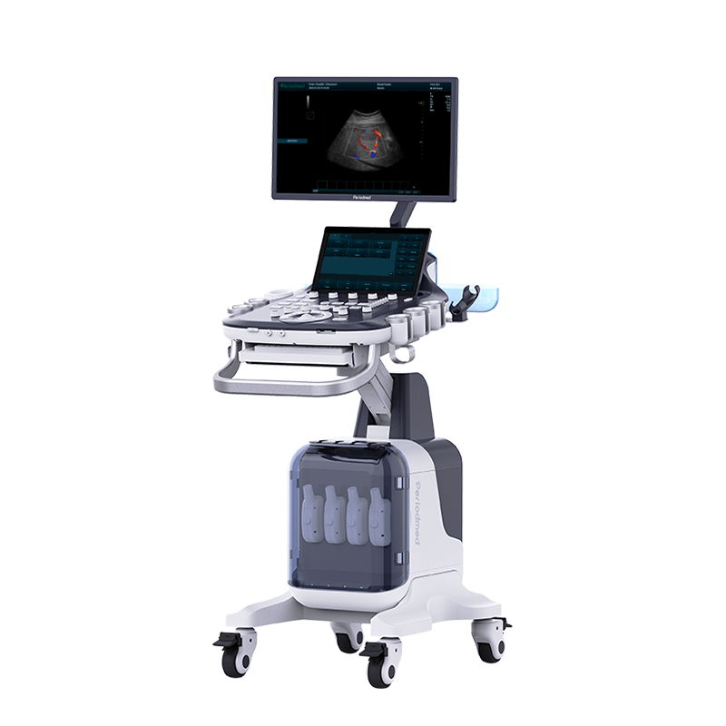
Featuring a 13.3-inch adjustable touch screen, four universal transducer ports and silent brake wheels, Revo T2 adapts flexibly to catheter lab environments. Integrated needle enhancement and multi-mode cardiac imaging optimize interventional scanning efficiency. Its stable high-precision imaging delivers consistent diagnostic quality for full-cycle structural heart interventional monitoring and post-operative assessment.
| Application | |||
| Generic | Obstetrics | Gynecology | Abdomen |
| Vascular | Small Parts | Urology | Thyroid |
| Cardiology | MSK | ||
| Image Formats | |||
| AVI | BMP | JPG | PNG |
| DICOM | |||
| Transducer Types | |||
| Convex probe | Linear probe | Phased array | |
| Volume array | Intracavity probe | Micro convex array | |
| Operating Mode | |||
| B | 2B | 4B | CW |
| M | CF | B/M | PW |
| PDI | PIP | Trapezoid | Panorama |
| 3D/4D(Optional) | Wide view (Optional) | Picture in Picture | Color M |
| Anatomy M | Directional Power Doppler Imaging | TDI(Optional) | Needle Enhance (Optional) |
Contrast Enhance(Optional) | Elastography (Optional) | SMV(Optional) | |
| System Architecture | |||
| Elements: 64/128/160/192 | Frequency Range: 2.0-22.0 MHz | Memory:8G | Hard Drive:256G |
| Dynamic Range: ≥120db | 13.3 Inches touch screen | Resolution:1920*1080 | Beam forming 16 |
| Frequency Range: 2.0-22.0 MHz | 23.8 Inches High Resolution LED Monitor | ||
| Physical Specs | |||
| 23.8 Inches High Resolution LED Monitor | Magnetic latch closure | Built-in speaker | Brightness and contrast adjustable |
| Languages | |||
| English | Arabic | French | Indonesian |
| Italian | Portuguese | Spanish | Chinese |
| German | Russian | ||


 EN
EN CN
CN