Tel/Whatsapp: +86 19352847010
Email: [email protected]

- Product
Ultrasound System
Life Information Monitoring System
Infusion & Syringe System
Solution
- About Us
- Media & Events
- Service
- Contact Us
Tel/Whatsapp: +86 19352847010
Email: [email protected]
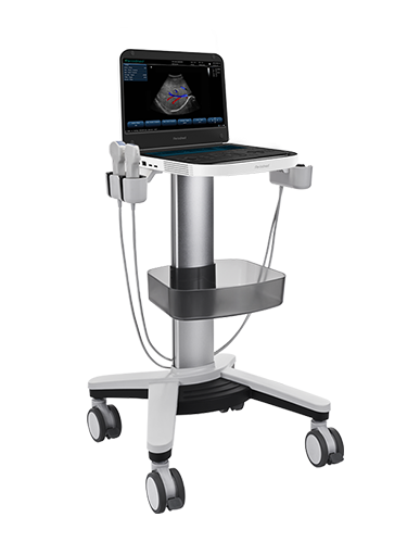
Integrated Hemodynamic Evaluation | Revo 9

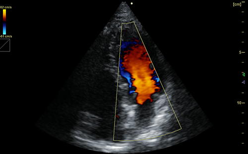
Revo 9 high-end ultrasound system delivers accurate cardiopulmonary ultrasound POCUS for comprehensive bedside hemodynamic assessment. Equipped with advanced PrismX™ and PureSight™ imaging technologies, it captures clear lung and cardiac tissue details. It integrates core clinical indicators to support systematic bedside monitoring, empowering clinicians to evaluate cardiopulmonary function and fluid status in one streamlined scan.
Specialized for POCUS for pulmonary congestion detection, Revo 9 precisely identifies and quantifies lung ultrasound B-lines indicative of interstitial edema and heart failure. Intelligent speckle reduction eliminates artifacts, ensuring reliable visualization of subtle extravascular lung water changes. This high-sensitivity lung assessment enables early recognition of pulmonary congestion before clinical symptoms become apparent.
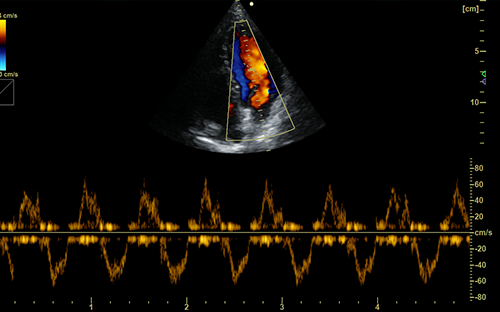
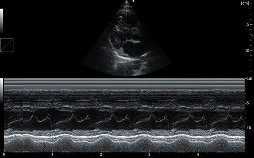
Revo 9 supports professional IVC ultrasound measurement and venous excess ultrasound score assessment for systemic venous congestion grading. It evaluates IVC collapsibility and abdominal venous hemodynamics to calculate standardized VExUS scores. Clinicians can accurately distinguish fluid overload, venous stasis and cardiac congestion, providing objective evidence for targeted decongestion and volume management strategies.
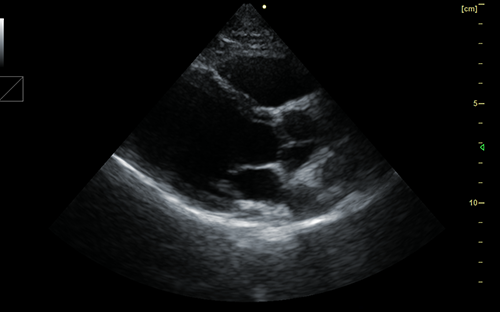
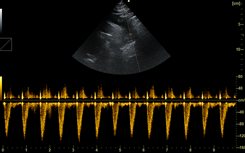
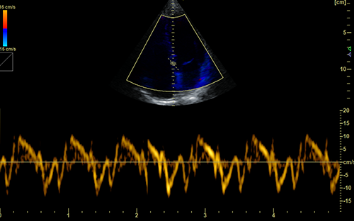
Optimized for LVOT-VTI hemodynamic monitoring bedside, the system performs accurate left ventricular outflow tract velocity-time integral measurement. Real-time Doppler and tissue tracking technologies reflect cardiac stroke volume and forward blood flow dynamically. It reliably assesses fluid responsiveness and cardiac output changes, guiding individualized fluid resuscitation and hemodynamic stabilization in critical care settings.
Revo 9 unifies B-lines lung scanning, VExUS grading and LVOT-VTI quantification to realize all-in-one cardiopulmonary hemodynamic evaluation. Its wide dynamic range and multi-mode imaging adapt to complex critical patient conditions. With DICOM data archiving and intelligent optimization functions, it supports continuous trending monitoring and standardized clinical decision-making for critical care and anesthesia departments.
| Application | |||
| Generic | Obstetrics | Gynecology | Abdomen |
| Vascular | Small Parts | Urology | Thyroid |
| Cardiology | MSK | ||
| Image Formats | |||
| AVI | BMP | JPG | PNG |
| DICOM | |||
| Transducer Types | |||
| Convex probe | Linear probe | Phased array | Micro convex array |
| Volume array | Intracavity probe | ||
| Standard Mode | |||
| B | 2B | 4B | CW |
| M | CF | PDI | PW |
| DPDI | Picture in Picture | Trapezoid | Panorama |
| Color M Mode | Anatomy M | TDI(Optional) | Wide View(Optional) |
| 3D/4D Mode(Optional) | Needle Enhance Mode(Optional) | Contrast Enhance Mode(Optional) | SMV(Optional) |
| Elastography(Optional) | |||
| System Architecture | |||
| Elements: 64/128/160/192 | Frequency Range: 2.0-22.0 MHz | Channel:64 | Hard Drive:256G |
| Dynamic Range: 36-120db | 13.3 Inches touch screen | Resolution:1920*1080 | Beam forming 16 |
| Memory:8G | 23.8 Inches High Resolution LED Monitor | ||
| Languages | |||
| English | Arabic | French | Indonesian |
| Italian | Portuguese | Spanish | Chinese |
| German | Russian | ||


 EN
EN CN
CN